Description
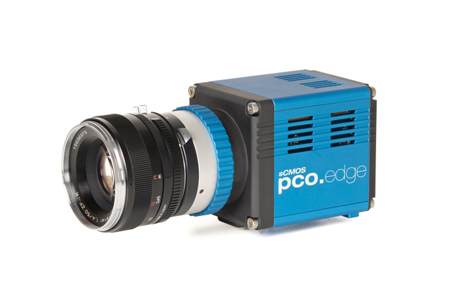
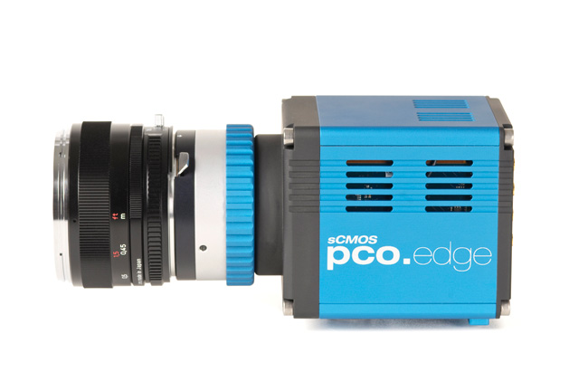
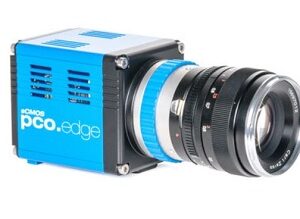
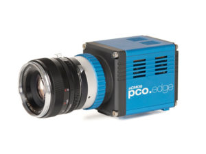
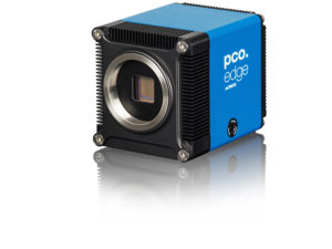
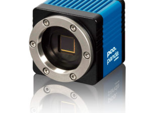
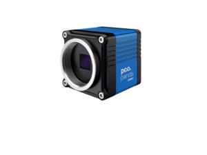
Innovations aren’t always about having that one big new idea. Unique technology also comes from evolution, combining existing and new technology. When PCO’s tried and trusted pco.edge series pools forces with modern back illuminated (bi) sensor technology, the result is the pco.edge 4.2 bi.
The adjustable cooling system allows the use of air or water to cool the sensor down to -25 °C. At this temperature, the dark current is reduced to 0.2 e-/pixel/s.
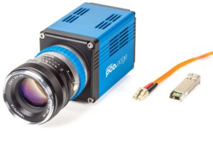
According to your application you can choose an input window specialized either for the UV or VIS range. High resolution and 6.5 x 6.5 μm² pixel size guarantees high-quality images with quantum efficiency up to 95 %. The pco.edge 4.2 bi incorporates a powerful USB 3.1 interface complementing its performance. Use the pco.edge 4.2 bi sCMOS camera system with the latest software from PCO.
Key Features:
- back illuminated 16 bit sCMOS sensor technology
- adjustable down to -25 °C air & water cooling
- high resolution 2048 x 2048 pixel
- pixel size: 6.5 x 6.5 µm²
- quantum efficiency up to 95 %
- maximum frame rate 40 fps @ full resolution
- dynamic range 26667 : 1
- lowest dark current (typ.) 0.2 e-/pixel/s
- rolling shutter
- exposure times from 10 μs to 20 s
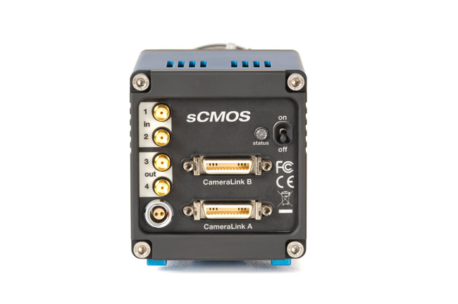
- USB 3.1 Gen1 interface
Quantum Efficiency Curve:

View/Download the datasheet here.
Application Areas:
- GSDIM
- Time Lapse 3-D Measurements
- PALM
- STORM
- SPIM
- SIM
- spinning disk confocal microscopy
- FRET
- FRAP
- EOS 3D
- adaptive optics
- solar astronomy
- fluorescence spectroscopy
- bio- & chemi-luminescence
- high content screening
- ophtalmology
- flow cytometry
- biochip reading
Application Examples:
Modular Wafer Inspection with pco.edge 5.5
 Silicon carbide (SiC) is a wide bandgap semiconductor, which is especially used for high-power, high-temperature and high-frequency devices due to its high energy efficiency. Despite great improvements in the material quality of SiC substrates and epitaxial wafers within the last years, critical defects like stacking faults (SFs) and basal plane dislocations (BPDs) can still lead to bipolar degradation and finally to complete failure of the device. Read more about this here.
Fluorensce Microscopy DNA Micro Arrays with pco.edge series
 Microarrays are versatile tools for high throughput screening. Nevertheless they are severely limited. Either the molecules are synthesized in-situ directly on the surface or in-vitro or in-vivo produced externally and then transferred onto the surface. In-situ synthesis shows low yield in terms of purity and restricts therefore the biomolecule probe length to ~50 bp for light-synthesized DNA (Affymetrics) or ~200 bp print-synthesis (Agilent), but allows up to millions of spots per array. Ex-array synthesis on the other hand provides high-purity molecules, but the (bio)synthesis and purification of these molecules is tedious, time consuming and expansive. Also the printing process takes time. Even if one spot can be made per second 100,000 spots will take more than a day. Therefore the idea arose why to copy microarrays. Why not make DNA, RNA and protein microarrays as high quality copies of a high quality original? It worked fine for text books and images. So why not apply it for DNA? Why not build a biomolecule copying machine? A biomolecule xeroxer? Read more about this here.
Time Lapse 3D measurements with pco.edge series
 Light sheet fluorescence microscopy has previously been demonstrated on a commercially available inverted fluorescence microscope frame using the method of oblique plane microscopy (OPM). In this paper, OPM is adapted to allow time-lapse 3-D imaging of 3-D biological cultures in commercially available glass-bottomed 96-well plates using a stage-scanning OPM approach (ssOPM). Read more about this here.
High Speed Florescence Microscopy with pco.edge series / pco.1600 / pco.sensicam
 The application shows that the mode and dynamics of trypanosome locomotion are a trait of life within a crowded environment. Using high-speed fluorescence microscopy and ordered micro-pillar arrays we show that the parasites mode of motility is adapted to the density of cells in blood. Read more about this here.
3D Flow Field Measurements with pco.edge 5.5/pco.dimax series
 Large-volume volumetric flow experiments and their results: The first of these, investigating a thermal plume at low velocities (up to 0.35 m/s) demonstrates the abilities and requirements to reach volume sizes up to and probably beyond one cubic mete Read more about this here.