Description
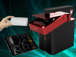
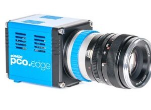
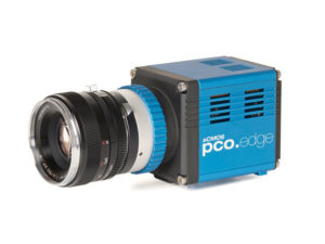
Abberior Instruments’ FACILITY platform offers you a workhorse instrument that boasts top-of-the-line superresolution STED and confocal imaging. It combines cutting-edge microscopy with unprecedented ease-of-use. Their software allows beginners to intuitively arrive at a top-notch image within three clicks, while also giving experts full control over the instrument. Microscopy has never felt better. ADAPTIVE ILLUMINATION (DYMIN, RESCUE, MINFIELD), Adaptive Optics, EASY3D STED, confocal & STED autofocus, spectral RAINBOW detection, and full AUTOALIGNMENT can all be included in the system.
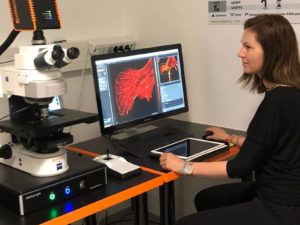
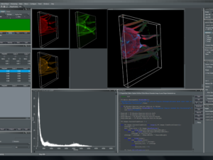
Abberior’s new software was designed from scratch to furnish a user interface that allows beginners to intuitively arrive at a top-notch STED image within three clicks, while it triggers a steep learning curve and allows expert users full control over the instrument. The unprecedented workflow outshines everything you have seen before.
White paper: Guided STED nanoscopy enables super-resolution imaging of blood stage malaria parasites
Jan-Gero Schloetel, Jörn Heine, Alan F. Cowman & Michał Pasternak
Scientific Reportsvolume 9, Article number: 4674 (2019)
Malaria remains a major burden world-wide, but the disease-causing parasites from the genus Plasmodium are difficult to study in vitro. Owing to the small size of the parasites, subcellular imaging poses a major challenge and the use of super-resolution techniques has been hindered by the parasites’ sensitivity to light. This is particularly apparent during the blood-stage of the Plasmodium life cycle, which presents an important target for drug research. The iron-rich food vacuole of the parasite undergoes disintegration when illuminated with high-power lasers such as those required for high resolution in Stimulated Emission Depletion (STED) microscopy. This causes major damage to the sample precluding the use of this super-resolution technique. Here we present guided STED, a novel adaptive illumination (AI) STED approach, which takes advantage of the highly-reflective nature of the iron deposit in the cell to identify the most light-sensitive parts of the sample. Specifically in these parts, the high-power STED laser is deactivated automatically to prevent local damage. Guided STED nanoscopy finally allows super-resolution imaging of the whole Plasmodium life cycle, enabling multicolour imaging of blood-stage malaria parasites with resolutions down to 35 nm without sample destruction.
To read more, click here
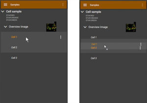
Tree View
All images are grouped hierarchically in a “tree view”, where context between measurements is kept from the sample level over large overviews down to final images. This way, STED and confocal measurements can always be spatially related to the greater environment of the sample, and you can switch between different modes at the push of a button. There is no need to zoom out to find a good area, followed by zooming in, adjusting settings such as pixel size and line frequency, taking an image, zooming out again, re-adjusting pixel size… all this is one click on the FACILITY. On top, the tree view supports drag and drop (right) to transfer settings to other images. Once you have found good settings for your sample, they can be applied lightning fast to all images, in this case from a measured “Cell 1” to a new region of interest called “Cell 2”
Tree View Supports Drag and Drop
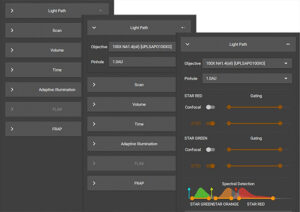
The FACILITY software supports new and advanced users alike. After entering the dyes that are in your sample, the machine knows everything about excitation wavelengths, detection bands, STED settings, etc. From there, you simply select a region you want to image and the first scan will give you a good superresolution measurement. After this, it’s a breeze to optimize settings.
All functions are grouped in convenient tabs that can be escalated to adapt to different user levels. In the example above, Light Path controls are unfolded, first to show basic objective and pinhole controls, while a more advanced user can move one escalation higher to manipulate gating and detection settings. This way, you only see what you need in the moment, cluttered screens with hundreds of buttons are avoided, and new users don’t feel overwhelmed, while advanced users are in full control.
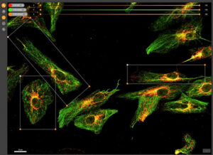
Free selection of ROIs
The FACILITY software allows tiled scans, and in every scan you can select an arbitrary number of regions of interests (ROIs, corresponding to items in the tree view, see above). Every ROI itself can serve as an overview again, meaning that you can easily climb down from sample level overviews to detailed superresolution images, while spatial context is saved at all times. Of course, the FACILITY supports arbitrarily rotated ROIs, even in STED mode!