 The INFINITY platform is the most customizable platform for all things microscopy. Whether it’s STED and confocal microscopy, intravital microscopy, material science or optical trapping, the INFINITY is always up to the challenge. Just tell us what you need and we will build you a customized, continuously upgradable system specialized for your research
Description
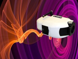
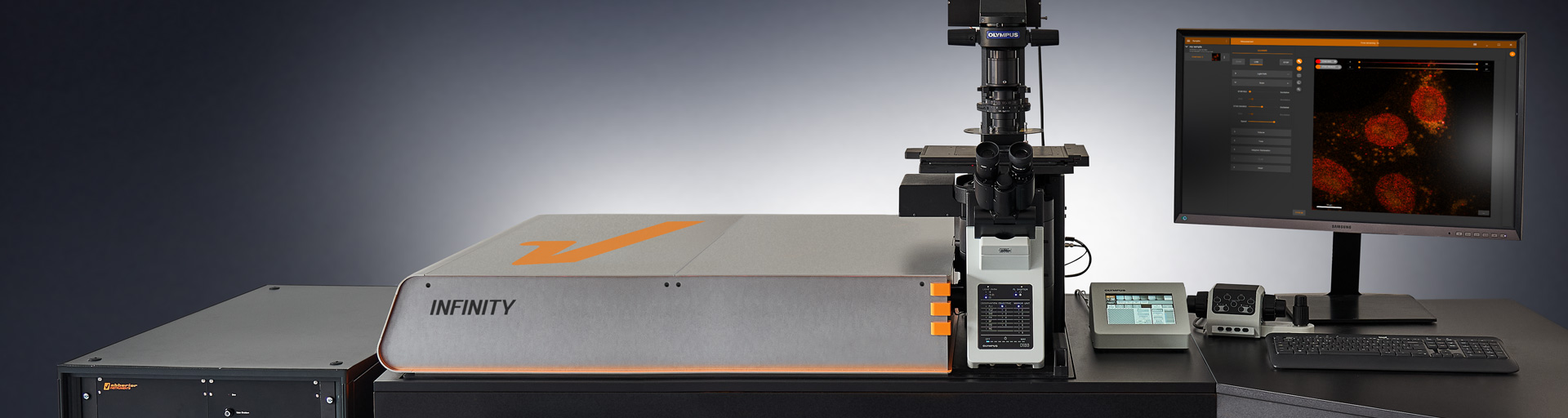
Abberior Instruments Infinity
The INFINITY is a highly flexible open platform that can be extended and upgraded, even years after purchase. It can be combined with all expert DEVICES and any custom developments to form a truly unique microscope that suits all your imaging needs. Get in touch with us and we will design and manufacture a tailor-made instrument that leaves nothing to be desired.
To protect your investment, we guarantee that your INFINITY grows with your science. And it grows with us. Your system could be a totally different machine a few years from now, because we at abberior make sure that everything we develop in the past, present or future can be retrofitted into your microscope. This way, you always stay at the forefront of science.
MODULES FOR STED / RESOLFT
- Matrix Detector
- Adaptive Illumination
- RESCue STED
- ADAPTIVE OPTICS
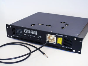
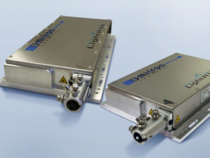
- STED Lasers
- EASY3D STED
- Autoalignment
- Autofocus
- Rainbow Detection
- FLIM
- Excitation Lasers
- Accessories
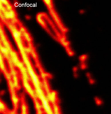
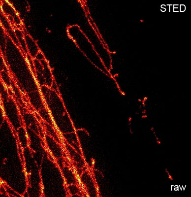
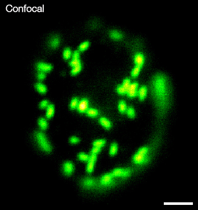
See below for a wide range of applications comparing Abberior Infinity STED with standard Confocal Microscopy
3-color STED imaging: Active zones at the Drosophila larval neuromuscular junction immunostained for Bruchpilot and two other proteins. Two superresolution channels (magenta, yellow) using a 775nm STED laser & one superresolution channel using a 595nm STED laser. Samples by M. Lenz & M. Landgraf (University of Cambridge, UK).
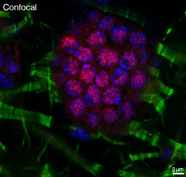
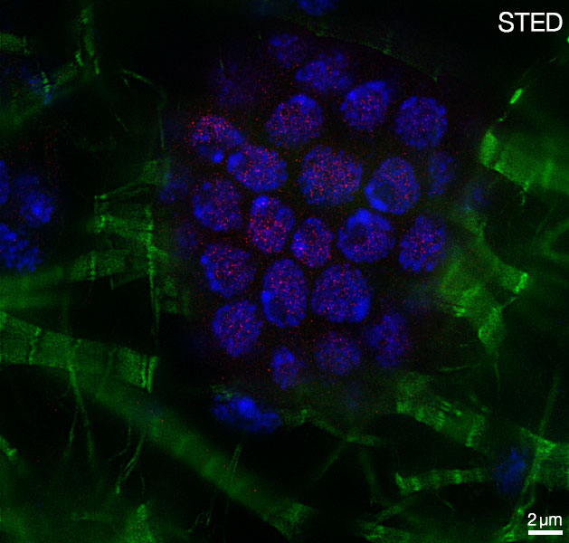
3-color RESCue STED images using both pulsed STED lasers @775nm and @595nm. Labelled structures: Nuclear pore complex (green, nup153, Oregon Green 488), Vimentin (white, Abberior STAR 635P) and lamin (red, Abberior STAR 580).
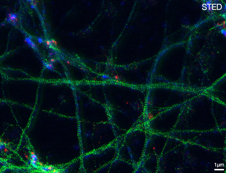
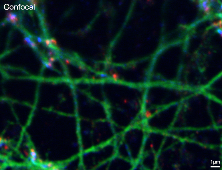
3-color STED image of primary hippocampal neurons. Please note the characteristic ~190 nm beta II spectrin periodicity along distal axons (green) which is only visible in the STED image. Labelled structures: beta II Spectrin (green, Abberior STAR635P), Bassoon (red, Abberior STAR580), Actin cytoskeleton (blue, Phalloidin, Oregon Green 488). Imaged with Abberior Expert Line with 595nm and 775nm STED laser. Sample was prepared by Elisa D’Este @ MPIBPC, Göttingen.
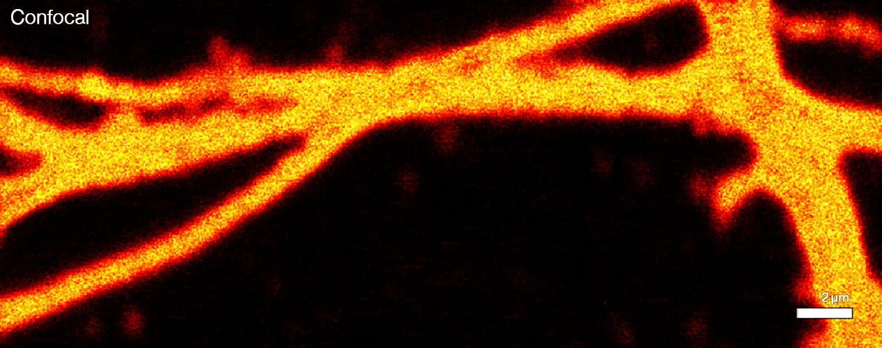
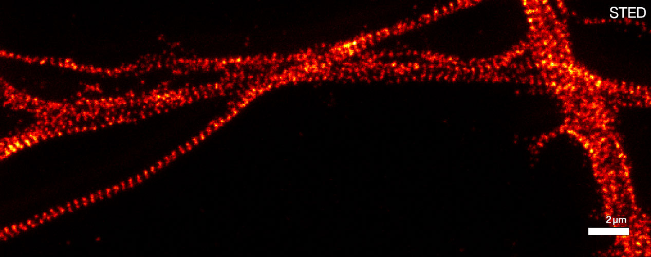
Primary hippocampal neurons (22 days in vitro) show the characteristic ~190 nm betaII spectrin periodicity along distal axons. Imaged with Abberior Expert Line @ MPIbpc by Elisa D’Este.
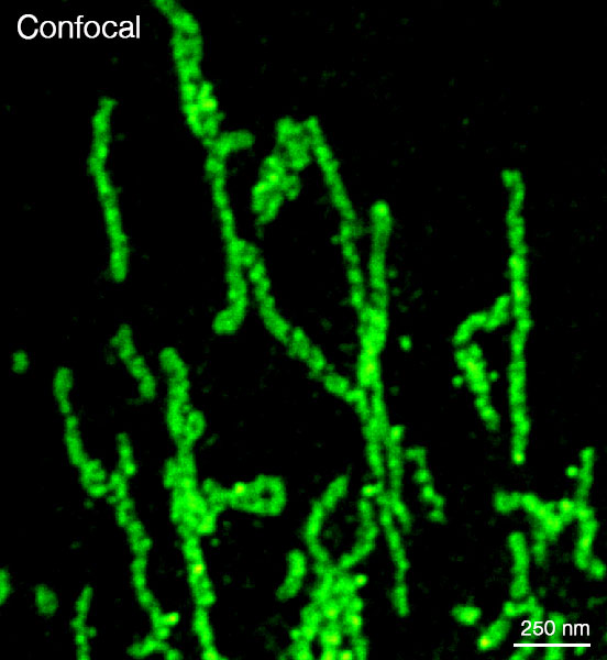
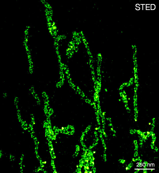
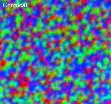
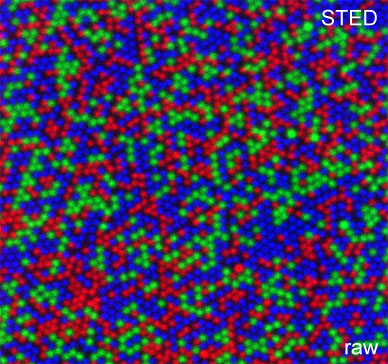
Mitochondrial protein Tom20 labeled with Alexa488 (in Vero cells). Shown is RAW DATA.
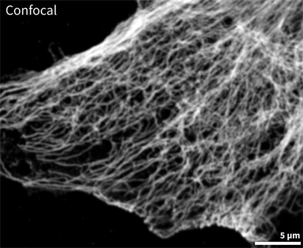
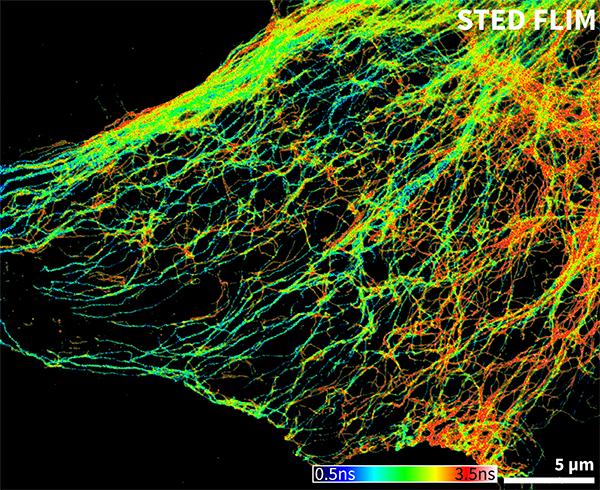
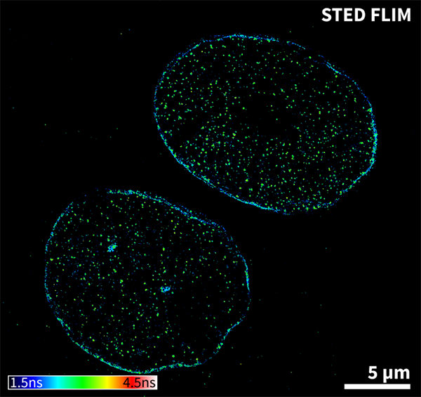
Confocal and STED FLIM image of mammalian cells labelled with tubulin/ Atto647N and vimentin/ Abberior STAR 635P.
STED reveals much smaller clusters of Cut protein and fine actin bundles. Samples were prepared by J. Rehman, Abberior GmbH / Dr. H. R. Shcherbata, MPI for Biophysical Chemistry, Göttingen, Germany.
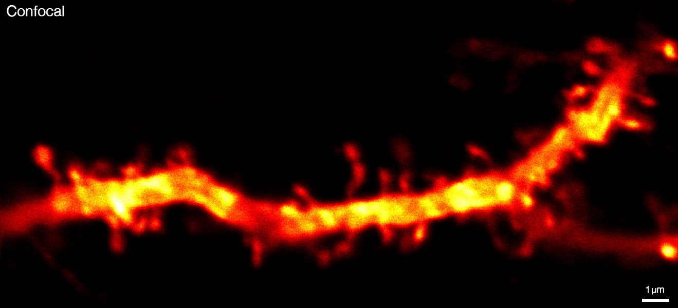
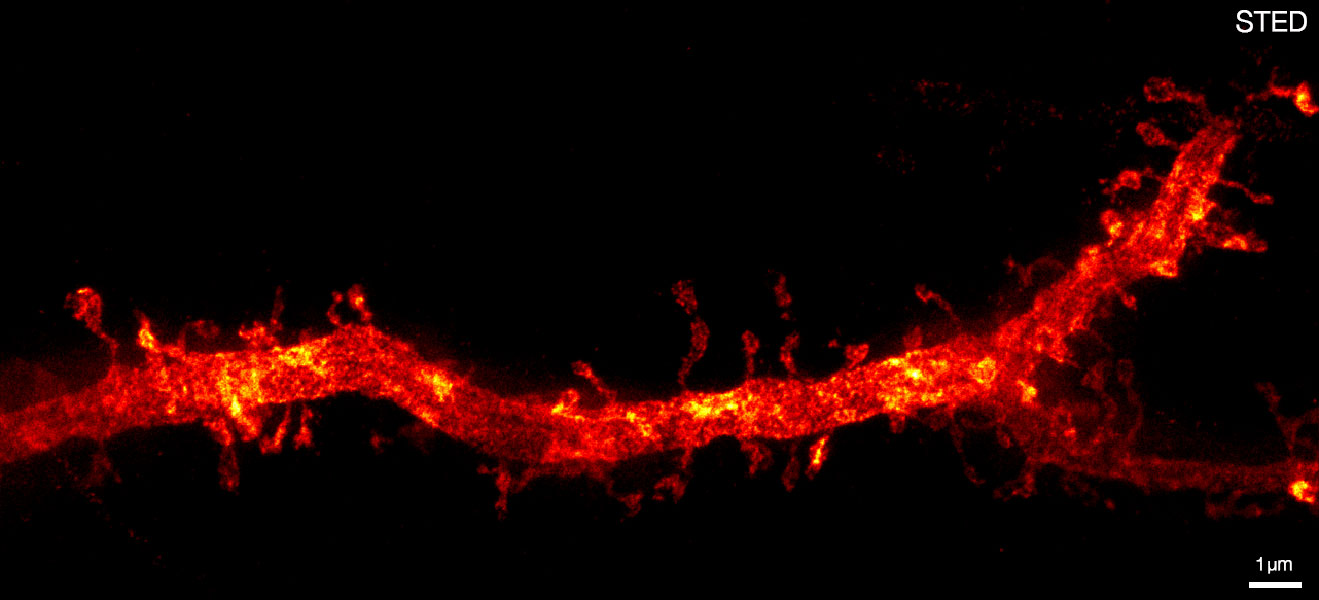
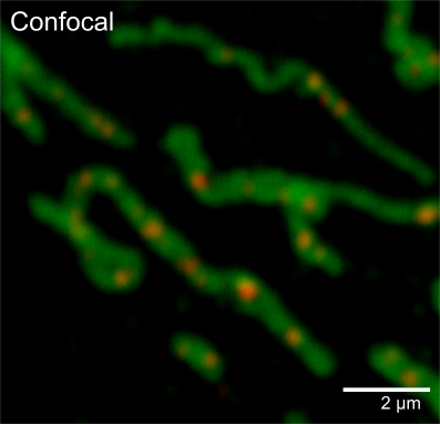
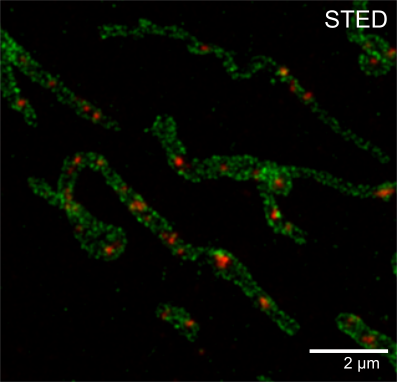
Confocal and 2D STED imaging of dendrites in brain slices. GFP-tagged proteins were expressed and immunolabelled using primary antibodies against GFP and secondary antibodies coupled to Abberior STAR 635P. Sample was prepared by O. Kaplan and H. Kawabe @ MPI of Experimental Medicine, Göttingen, Germany.
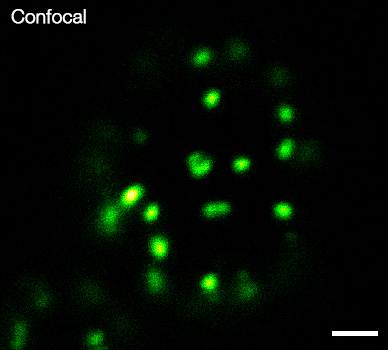
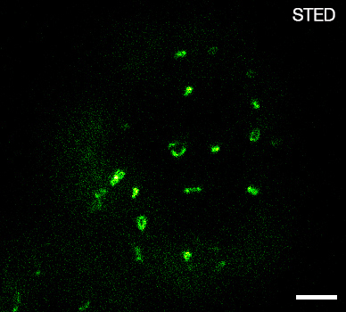
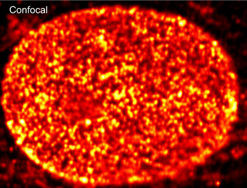
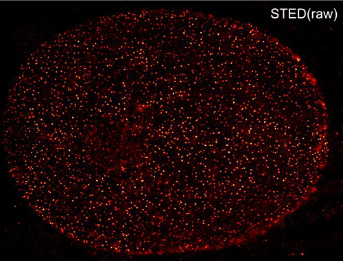
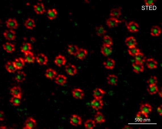
Nuclear pore complex protein (nup153). Resolution (STED): < 25 nm.
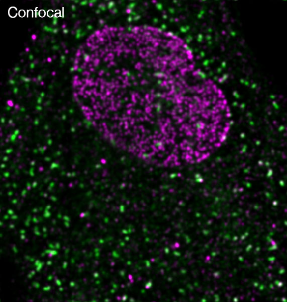
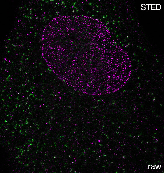
Nuclear pore complex (nup153, violett) and peroxisomal protein (PMP70, green) Resolution STED < 25 nm.
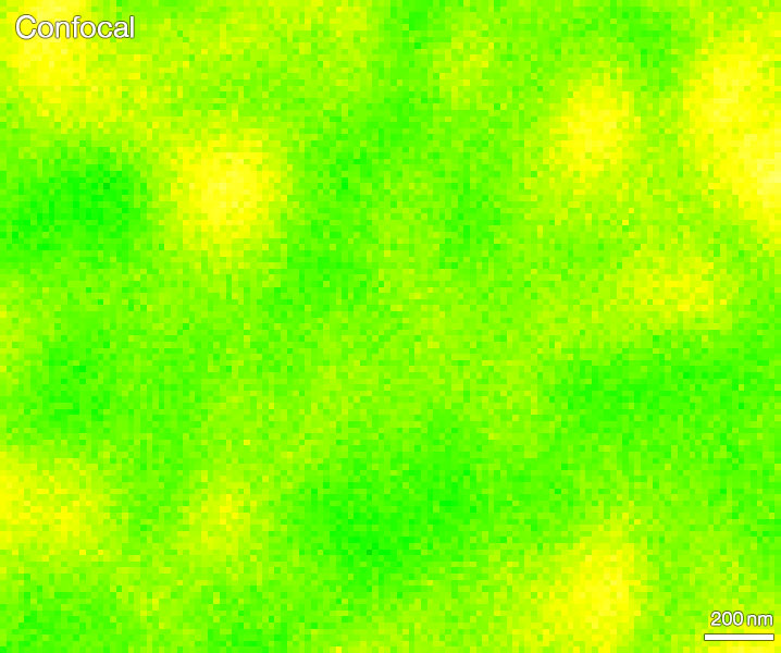
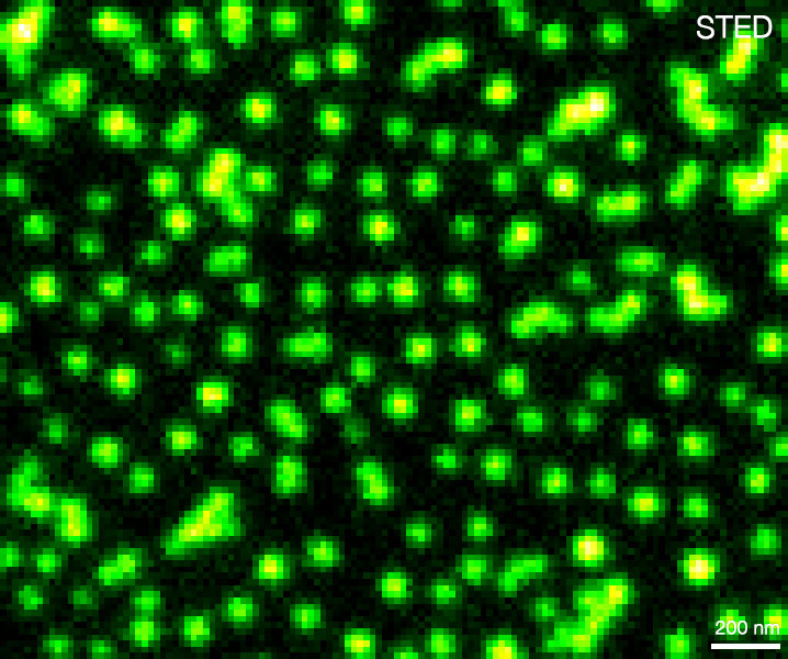
Mixture of 3 fluorescent bead species with 3 different colors (200nm diameter) Resolution STED < 35nm (green) and STED < 25nm (red, blue).
Confocal FLIM and STED FLIM image of mammalian cells labeled with antibodies against lamin/ Atto647N and DNA/ Abberior STAR 635P.
Confocal and STED image of green fluorescent beads. Measured with our Expert Line @ 595nm pulsed STED.
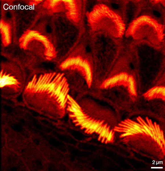
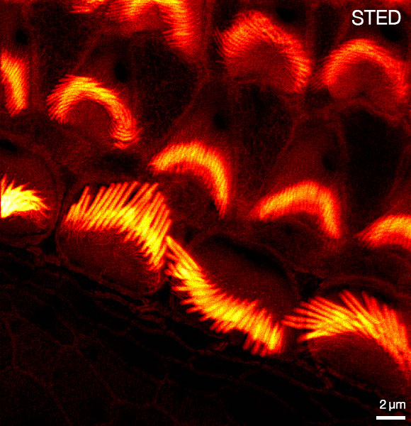
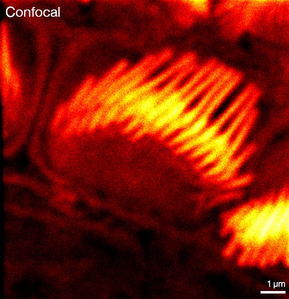
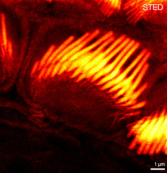
Actin stain of mouse inner ear hair cells using Abberior STAR RED Phalloidin. Samples were prepared by Dr. Christian Vogl, InnerEarLab, UMG Göttingen, Germany.
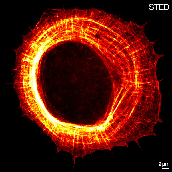
Actin organization in a primary astrocyte at 1 day in vitro. Imaged with Abberior Expert Line @ MPIbpc by Elisa D’Este.

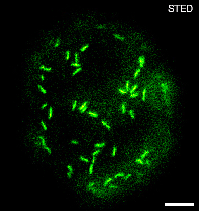
Bruchpilot (sample courtesy of Prof. Sigrist, FU Berlin) Resolution STED < 25 nm.
Citrine labeled eisosomes in living yeast cells. RAW DATA is shown. Scalebar, 1 µm (sample courtesy of Prof. Dr. S. Jakobs, University Medical Center Gottingen).
Actin stain of mouse inner ear hair cells using Abberior STAR RED Phalloidin. Samples were prepared by Dr. Christian Vogl, InnerEarLab, UMG Göttingen, Germany.
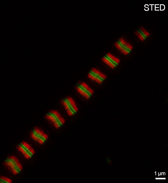
STED and confocal images of two sarcomeric proteins labeled with Abberior STAR 580 and Abberior STAR RED. RAW DATA is shown. Sample courtesy of Prof. Dr. M. Gautel, King’s College, London.
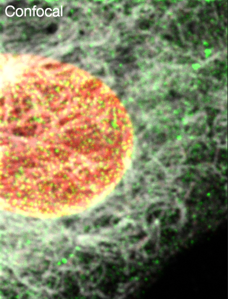
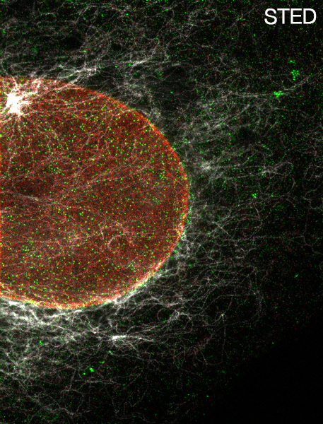
Mitochondria labeled with antibodies against Tom20 and dsDNA. Shown is RAW DATA.
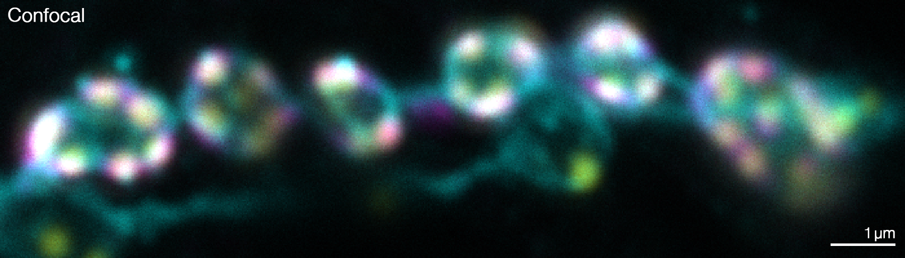
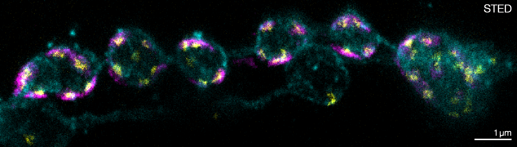
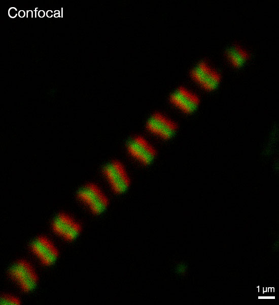
Two subunits of the nuclear pore complex were immunolabelled using antibodies against gp210 and antibodies with multiple specificities (PAN4/5) and secondary antibodies coupled to Abberior STAR 580 and Abberior STAR 635P. Note that gp210 is localized in an eightfold symmetric structure at the rim of the nuclear pore complex.
YFP labeled eisosomes in living yeast cells. RAW DATA is shown. Scalebar, 1 µm (sample courtesy of Prof. Dr. S. Jakobs, University Medical Center Gottingen).