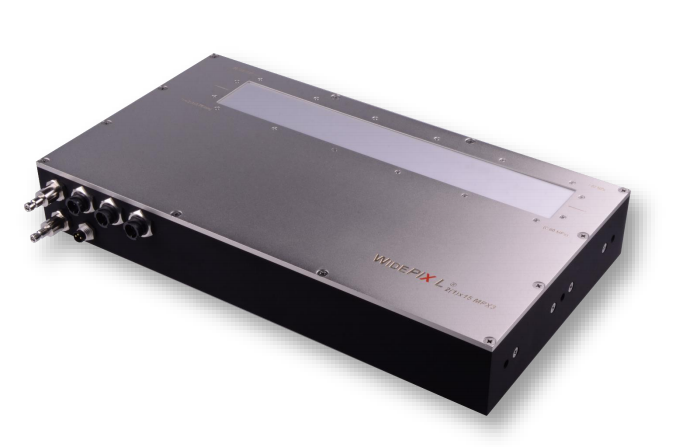
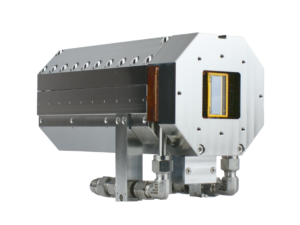
Very large area industrial x-ray spectral imaging camera with edge-less sensor technology with speed up to 50 fps and a size of 256 x 3840 pixels. Can be used for non-destructive testing, small animal spectral imaging, art authentication and mining and geology applications.
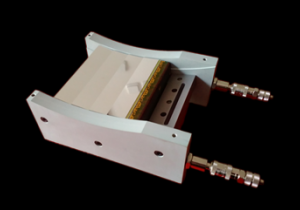
WidePIX 2(1)x15 – MPX3 camera consists of either 2×15 or 1×15 Medipix3 devices. Each pixel has two integrated 12-bit digital counters and two energy discrimination thresholds. Both counters can be joined to a single 24-bit counter providing enhanced dynamic range. The camera can be constructed with Si or CdTe edgeless sensor tiles. The edgeless sensor technology allows placing all tiles tightly together from all sides. Thus the whole imaging area of the camera is fully sensitive to the radiation – there are no gaps between the tiles in the image. The camera is made robust to address industrial users.
The WidePIX 1×15 – MPX3 camera offers built-in Time-Delayed-Integration (TDI) for scanning in industrial applications. It is very advantageous for continuous scanning. WidePIX 2(1)X15 – MPX3 is available with Si and CdTe sensors for imaging of the soft and hard X-ray spectra, respectively.
The minimum detectable energy is typically 3 keV or 5 keV in case of Si or CdTe sensor, respectively. The intrinsic spatial resolution of the camera is defined by pixel size which is 55 µm. The pixels situated on the border of tiles are 2 times larger in one direction. The corner pixels of tiles are 2 times larger in both directions.
Key Features of WidePIX 2(1)X15:
Sensor Material: Si or CdTe
Sensor Thickness: 300 μm and 500 μm for Si; 1 mm for CdTe
Sensitive Area: 14 mm x 211.2 mm (1×15 tiles) and 28 mm x 211.2 mm (2×15 tiles)
Number of Pixels: 256 x 3,840 (1×15 tiles) and 512 x 3,840 (2×15 tiles)
Pixel Pitch: 55 μm
Resolution: 9 lp/mm
Readout Speed: 50 (1×15 tiles) and 20 (2×15 tiles) frames/s
Photon Counting Speed: up to 3 x 10⁶ photons/s/pixel
Continuous readout (DTDI): Yes, hardware based (1×15 tiles)
Threshold Step Resolution: 0.1 keV
Threshold per pixel: 1, 2 or 8 (configurable)
Energy Resolution: 0.7-2.0 keV (Si) and 1.2-3.6 (CdTe)
Min Detectable Energy: 2.5 keV (Si) and 5 keV (CdTe)
Readout Chip: Medipix3
Pixel Mode of Operation: Counting in Single Pixel Mode (SPM) or Charge Summing Mode (CSM)
Non-Destructive Testing (NDT) is inspection, test, or evaluation of materials, components or assemblies for discontinuities, or differences in characteristics without destroying the serviceability of the sample. Standard radiographic X-ray imaging provides a black and white intensity or density image of the inspected sample where defects, impurities or cracks are observed if the resolution and the signal over the noise of the image is appropriate. The spectral NDT X-ray imaging provides based on photon counting provides additional material information of the samples together with a superior contrast and high spatial resolution. The spectral material information is used to discriminate different materials that can be used to identify the materials of interest or to calculate their amount in the sample. Find out more about this application here.
Small Animal Spectral Imaging
Cancer research, bio-mechanics, and drug testing are just a few examples of where X-ray imaging contributes to research in biology and medicine. New photon counting detectors represent a serious advancement for these applications, compared to previously used synchrotrons. The energy sensitivity of modern cameras opens better possibilities to identify individual types of tissue. That has important consequences in various industries, for example cancer research, where the tumour tissue can be better distinguished from the healthy one. Find out more about this application here.
Art Authentication
Study and characterisation of art pieces, namely paintings, using X-ray imaging is becoming an increasingly important area. It is useful for galleries, museums and collectors to improve conservation and preservation methods. It is important for art buyers to reliably authenticate the works. The advanced X-ray imaging techniques provide detailed data for insurance companies to assess risks involved in transportation of art. The spectral imaging capability of Advacam’s detectors enables identification of different pigments based on their spectral responses. A “colour” X-ray image is then created where the colours are associated to different pigments identified in the painting. The recognition of pigments even in invisible lower layers can serve as an important clue in the process of art authentication. Find out more about this application here.
Mining and Geology
Resources mined from Earth fuel our society. However, efficient, eco-friendly mining is a must if we want to keep the economy and society sustainable. Cheap, fast exploration for mineral resources is required. Geology is a tool to understand our planet and also to help find resources still undiscovered. The energy sensitive photon counting detectors can help in this area thanks to the option of material identification in images. Drill cores from exploration bore holes can be now analysed in-situ identifying different minerals and providing immediate feedback for further exploration. Online monitoring of technological processes during mineral processing is another very important area where X-ray material resolved imaging plays an important role. It helps to increase efficiency of processing and reduce energy consumption leading not only to reduced costs, but also to lower environmental effects. Find out more about this application here.
Advacam Technology
The leading detector technology, which Advacam uses for its products and solutions is based on Medipix hybrid pixel detectors. These devices were developed within international collaboration of universities and research laboratories lead by team at CERN during past 20 years. Advacam team’s members have been part of the Medipix Collaboration from it inception and have been contributing to the technology.
Photon Counting Technology
Advacam’s imaging cameras are direct conversion single photon counting pixel detectors that represent the cutting edge of current radiation imaging technology. The term “single photon counting” means that every single photon of X-ray radiation detected in individual pixel is processed and counted. The technology brings two major advantages in comparison to the conventional X-ray imaging – high contrast together with sharp images and spectral information of X-rays that allows material specific information to be displayed in colors.
In the direct conversion cameras each pixel of the semiconductor crystal is directly connected to the complex CMOS circuit using a conductive solder bump. In the indirect conversion cameras a scintillation layer is attached on top of a photodiode. The photodiodes manufactured on a simple CMOS circuit that enables fine pixel sizes.
Illustrative comparison of a single pixel of a direct conversion and indirect conversion cameras.
The term direct conversion refers to immediate conversion of the X-rays into electric charge within the semiconductor crystal. The principle is contrary to the conventional indirect conversion where the X-rays are first converted into visible light in the scintillation layer that subsequently is converted into electric charge in the photodiodes.
Illustration of the operation principles in a single pixel between the direct and indirect conversion cameras.
The photon counting principle of detection eliminates all other sources of noise that are present in CCD or flat-panel based cameras. This leads to considerably better signal-to-noise ratio and therefore detectability of more details in images. The images sharpness or the actual spatial resolution of the captured image is defined by the electric charge in the CMOS readout. Even thought the pixel size of of the direct converting cameras is larger than that of the conventional indirect conversion cameras, the signal of the detected X-rays is better focused into the pixels. The typical size of a direct conversion pixel ranges from few millimeters to tens of micro meters where Advacam represents the highest pixel density of the current industrial X-ray cameras with 55 um pixel size. The video below describes the differences between conventional indirect conversion, direct conversion charge integrating and photon counting cameras. It summarises the major differences in the captured image quality in terms of spatial resolution, image noise and material discrimination.
The energy sensitivity is as important advancement of the imaging technology as was the colour photography and film. Contrary to regular X-ray imaging cameras, the photon counting cameras can discriminate or even directly measure energy (wavelength) of incoming photons. Since each element of the sample has different X-ray attenuating properties, it is possible to estimate material composition of the sample if the energy of the photons is measured. The spectral sensitivity offers major improvement over the conventional X-ray imaging cameras.
Digital Time Delayed Integration
Digital Time Delayed Integration (DTDI) allows continues scanning of the inspected samples without stitching of individual images that cannot fit into the field of view of the camera. The high quality and resolution image is formed as the sample moves across the field of view of the camera. The scanning speed of the camera or the movement of the sample (for example on a conveyor belt) can be up to tens of meters per second.