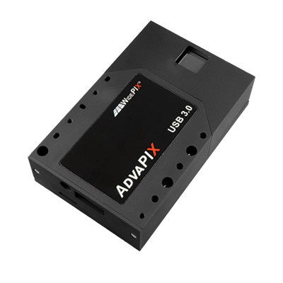
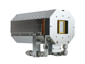
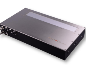
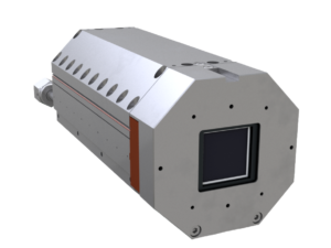
The AdvaPIX camera is designed with special emphasis to performance and versatility that is often required in a scientific experimental work. The camera module contains a single Timepix device with a fast parallel readout. It allows a sustainable frame rate of up to 1700 frames per second that makes it optimal for spectral imaging and ion detection. Advanced data processing algorithms allow ion detection with spatial resolution of up to 200 nm.
AdvaPIX can be used in variety of applications such as fast radiography (X-rays, neutrons, ions), spectroscopic imaging (each pixel records the energy spectrum), particle tracking, Time-of-flight imaging, multilayer Compton camera (thin top sensor, thick bottom sensor). The sensor type and thickness can be customized and sensors can be also adapted for neutron imaging by deposition of converter layer (6LiF). The spatial resolution in some applications (thermal neutrons) can reach units of microns or even submicrometric level (ions).
Key Features of AdvaPIX:
Sensor Material: Si or CdTe
Sensor Thickness: 100 μm, 300 μm and 500 μm for Si; 1 mm for CdTe
Sensitive Area: 14 mm x 14 mm
Number of Pixels: 256 x 256
Pixel Pitch: 55 μm
Resolution: 9 lp/mm
Readout Speed: 1700 frames/s
Photon Counting Speed: Up to 3 x 10⁶ photons/s/pixel
Threshold Step Resolution: 0.1 keV
Energy Resolution: 0.8 keV (THL) and 2 keV (ToT)
Min Detectable Energy: 5 keV for X-rays
Readout Chip: Timepix
Pixel Mode of Operation: Counting, Time-over-Threshold, Time-of -Arrival
Non-Destructive Testing (NDT) is inspection, test, or evaluation of materials, components or assemblies for discontinuities, or differences in characteristics without destroying the serviceability of the sample. Standard radiographic X-ray imaging provides a black and white intensity or density image of the inspected sample where defects, impurities or cracks are observed if the resolution and the signal over the noise of the image is appropriate. The spectral NDT X-ray imaging provides based on photon counting provides additional material information of the samples together with a superior contrast and high spatial resolution. The spectral material information is used to discriminate different materials that can be used to identify the materials of interest or to calculate their amount in the sample. Find out more about this application here.
X-ray Diffraction
X-ray diffraction is analytical method based on inspection of crystalline structure of samples used in applications, such as metallurgy, mineralogy, powders, pigments, polymers, surface layers and strain mapping. The traditional X-ray diffraction uses monochromatic X-rays which make the apparatus large and slow. ADVACAM’s spectral detectors based on Timepix3 chip with high resolution makes the diffraction system fast and compact. The sample analysis can be performed 100 times faster compared to the conventional systems. Due to fast speed of the analysis large areas of the sample can be analysed by scanning. Find out more about this application here.
Mining and Geology
Resources mined from Earth fuel our society. However, efficient, eco-friendly mining is a must if we want to keep the economy and society sustainable. Cheap, fast exploration for mineral resources is required. Geology is a tool to understand our planet and also to help find resources still undiscovered. The energy sensitive photon counting detectors can help in this area thanks to the option of material identification in images. Drill cores from exploration bore holes can be now analysed in-situ identifying different minerals and providing immediate feedback for further exploration. Online monitoring of technological processes during mineral processing is another very important area where X-ray material resolved imaging plays an important role. It helps to increase efficiency of processing and reduce energy consumption leading not only to reduced costs, but also to lower environmental effects. Find out more about this application here.
Other Applications
ADVACAM offers cameras and solutions for multiple other application such as electron microscopy, crystallography, neutron imaging, charged particle tracking and dosimetry for cancer therapy. In addition, number of cameras are being used in the basic research, such as at CERN’s LHC accelerator, and for education of radiation and its properties in schools and universities. Find out more about this here.
Advacam Technology
The leading detector technology, which Advacam uses for its products and solutions is based on Medipix hybrid pixel detectors. These devices were developed within international collaboration of universities and research laboratories lead by team at CERN during past 20 years. Advacam team’s members have been part of the Medipix Collaboration from it inception and have been contributing to the technology.
Photon Counting Technology
Advacam’s imaging cameras are direct conversion single photon counting pixel detectors that represent the cutting edge of current radiation imaging technology. The term “single photon counting” means that every single photon of X-ray radiation detected in individual pixel is processed and counted. The technology brings two major advantages in comparison to the conventional X-ray imaging – high contrast together with sharp images and spectral information of X-rays that allows material specific information to be displayed in colors.
In the direct conversion cameras each pixel of the semiconductor crystal is directly connected to the complex CMOS circuit using a conductive solder bump. In the indirect conversion cameras a scintillation layer is attached on top of a photodiode. The photodiodes manufactured on a simple CMOS circuit that enables fine pixel sizes
Illustrative comparison of a single pixel of a direct conversion and indirect conversion cameras.
The term direct conversion refers to immediate conversion of the X-rays into electric charge within the semiconductor crystal. The principle is contrary to the conventional indirect conversion where the X-rays are first converted into visible light in the scintillation layer that subsequently is converted into electric charge in the photodiodes.
Illustration of the operation principles in a single pixel between the direct and indirect conversion cameras.
The photon counting principle of detection eliminates all other sources of noise that are present in CCD or flat-panel based cameras. This leads to considerably better signal-to-noise ratio and therefore detectability of more details in images. The images sharpness or the actual spatial resolution of the captured image is defined by the electric charge in the CMOS readout. Even thought the pixel size of of the direct converting cameras is larger than that of the conventional indirect conversion cameras, the signal of the detected X-rays is better focused into the pixels. The typical size of a direct conversion pixel ranges from few millimeters to tens of micro meters where Advacam represents the highest pixel density of the current industrial X-ray cameras with 55 um pixel size. The video below describes the differences between conventional indirect conversion, direct conversion charge integrating and photon counting cameras. It summarises the major differences in the captured image quality in terms of spatial resolution, image noise and material discrimination.
The energy sensitivity is as important advancement of the imaging technology as was the colour photography and film. Contrary to regular X-ray imaging cameras, the photon counting cameras can discriminate or even directly measure energy (wavelength) of incoming photons. Since each element of the sample has different X-ray attenuating properties, it is possible to estimate material composition of the sample if the energy of the photons is measured. The spectral sensitivity offers major improvement over the conventional X-ray imaging cameras.