Description
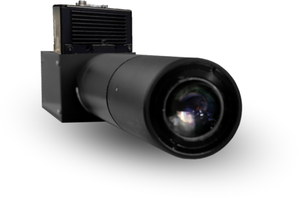
The ChromaLine 2.5D, All-in-Focus line scan vision system enables detailed inspection at high resolution of structurally complex, often highly reflective components on fast moving production lines. It performs image acquisition at up to 80,000 lines per second, and can be used to rapidly detect even the smallest features and defects in near real time.
ChromaLine principle of operation:
1) An incident white light line is imaged through a chromatic objective into a continuum of monochromatic linear images along the Z-Axis, thus providing a color-coded field along the optical axis (corresponding to the depth of focus).
2) When an object is present in this “coloured” field, a unique wavelength is perfectly focused at its surface and reflected into the optical system.
3) The backscattered light passes through a filtering pinhole and is imaged onto a CMOS line scan camera, thus creating a perfectly focused image of the object traveling through the entire range of depth-of-focus.
There are different optical heads available to allow for a wide range of resolutions and depths of the field.
Coaxial Acquisition
- Integrated Lighting
- Camera interface
- 4K Resolution
- Up to 80 KHz
Inspection vision systems have a dual role:
- Provide a high quality image of the surface of the sample with the desired magnification.
- Process the image to detect and analyze certain predefined features or textures.
As processing units become increasingly more powerful and sophisticated, the basic limitation most of the current vision inspection systems in use today lies in their very shallow depth of field or focus (a few tens of μm or less, depending on the numerical aperture). Focusing mechanisms are needed for viewing samples with large deviations in the depth of the surface. Chromatic Confocal Microscopy (STIL patent) is a technology that overcomes these limitations to provide a very large depth of focus of up to several mm. Hence these systems provide excellent image quality which are perfectly centred, even with very complex samples. This technology combines the advantages of colour coding and traditional confocal microscopes.
The confocal chromatic microscope consists of a slit illuminated by a polychromatic light source, a high quality chromatic lens, a beam splitter and a linear photo detector.
| MC2 | NanoView | MicroView | DeepView | SuperView |
|---|---|---|---|---|
| Line Length (mm) | 1.35 | 1.8 | 4 | 12.5 |
| Depth of Field (mm) | 0.1 | 0.5 | 2.6 | 2.0 |
| Working Distance (mm) | 4.6 | 10.1 | 47.8 | 12.5 |
| Pixel Size (μm2) | 0.4x0.41 | 0.54x0.54 | 1.2x1.2 | 3.82x3.82 |
| Slope angle (Deg) | +/-40 | +/-30 | +/-20 | +/-19 |
| Numerical Aperture | 0.7 | 0.5 | 0.35 | 0.32 |
Download data sheet here


