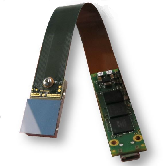
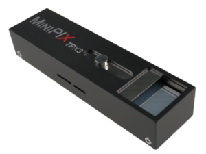
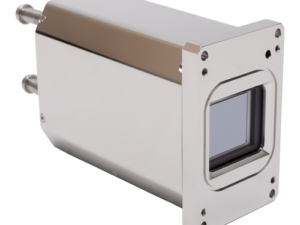
The MINIPIXTPX3F is miniaturized and low power radiation camera with particle tracking and imaging detector Timepix3 (256 x 256 square pixels with pitch of 55 μm). The MINIPIX TPX3F chip is equipped with sensor according to customer preference (standardl 300 μm thick silicon). The Timepix3 detector is position, energy and time sensitive: For each ionizing particle (e.g. X-ray photon) it digitally registers its position, energy, time of arrival and track shape – basically all information you can want. The other measures can be often calculated from the track shape (particle type, direction of flight, LET, charge …). The information on each detected particle is either read-out immediately (pixel mode) at maximal rate of 2.3 million hit pixels per second or accumulated in pair of images
(frame mode) and read-out later at maximal speed of 16 frames per second.
The MINIPIX TPX3F device is controlled via USB2.0 interface with standard µUSB connector. All major operating systems are supported (MS Windows, Mac OS and LINUX). The complex software PIXET PRO used for detector operation is provided for free. The extra software modules are available for special functions (e.g. coded aperture image reconstruction, Compton camera image and spectrum reconstruction, radiation field decomposition, networking of many devices …).
Spectral gamma ray imaging: scintigraphy or SPECT, radiography with isotopes.
Radiation monitoring: particle type sorting, spectroscopy, directional sensitivity …
Gamma camera: special shielded box and collimators available.
Compton camera: special software module available for image reconstruction.
Advacam Technology
The leading detector technology, which Advacam uses for its products and solutions is based on Medipix hybrid pixel detectors. These devices were developed within international collaboration of universities and research laboratories lead by team at CERN during past 20 years. Advacam team’s members have been part of the Medipix Collaboration from it inception and have been contributing to the technology.
Photon Counting Technology
Advacam’s imaging cameras are direct conversion single photon counting pixel detectors that represent the cutting edge of current radiation imaging technology. The term “single photon counting” means that every single photon of X-ray radiation detected in individual pixel is processed and counted. The technology brings two major advantages in comparison to the conventional X-ray imaging – high contrast together with sharp images and spectral information of X-rays that allows material specific information to be displayed in colors.
In the direct conversion cameras each pixel of the semiconductor crystal is directly connected to the complex CMOS circuit using a conductive solder bump. In the indirect conversion cameras a scintillation layer is attached on top of a photodiode. The photodiodes manufactured on a simple CMOS circuit that enables fine pixel sizes
Illustrative comparison of a single pixel of a direct conversion and indirect conversion cameras.
The term direct conversion refers to immediate conversion of the X-rays into electric charge within the semiconductor crystal. The principle is contrary to the conventional indirect conversion where the X-rays are first converted into visible light in the scintillation layer that subsequently is converted into electric charge in the photodiodes.
Illustration of the operation principles in a single pixel between the direct and indirect conversion cameras.
The photon counting principle of detection eliminates all other sources of noise that are present in CCD or flat-panel based cameras. This leads to considerably better signal-to-noise ratio and therefore detectability of more details in images. The images sharpness or the actual spatial resolution of the captured image is defined by the electric charge in the CMOS readout. Even thought the pixel size of of the direct converting cameras is larger than that of the conventional indirect conversion cameras, the signal of the detected X-rays is better focused into the pixels. The typical size of a direct conversion pixel ranges from few millimeters to tens of micro meters where Advacam represents the highest pixel density of the current industrial X-ray cameras with 55 um pixel size. The video below describes the differences between conventional indirect conversion, direct conversion charge integrating and photon counting cameras. It summarises the major differences in the captured image quality in terms of spatial resolution, image noise and material discrimination.
The energy sensitivity is as important advancement of the imaging technology as was the colour photography and film. Contrary to regular X-ray imaging cameras, the photon counting cameras can discriminate or even directly measure energy (wavelength) of incoming photons. Since each element of the sample has different X-ray attenuating properties, it is possible to estimate material composition of the sample if the energy of the photons is measured. The spectral sensitivity offers major improvement over the conventional X-ray imaging cameras.