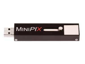
MiniPIX EDU is a miniaturised USB camera designed and priced for educational use. It brings modern nuclear technology of radiation imaging to the classrooms and let students discover the invisible world of ionizing radiation surrounding us. Students can explore the origin of different types of radiation and see how radioactive isotopes migrate in the nature and in the artificial environment of human houses, cities, industries. The same technology is used by NASA in space to monitor the radiation received by the astronauts.
MiniPIX EDU can record very small levels of radioactivity which is present everywhere. Students can see radioactivity of common materials and objects such as piece of granite, ash or paper bag from vacuum cleaner or face mask. They can explore variation of the air radioactivity during the day, hunt for cosmic muons and check their directions, see how altitude affects presence of radiation types. They can try to prepare their own (safe) radioactive source and try to construct the shielding against radiation it emits. They can check the laws of radioactive decay. Students can directly observe how different radiation types interact with matter and what happens then.
It is easy to plug the MiniPIX EDU device to the USB port of your PC and start the software. Using the proprietary RadView radiation visualisation software fascinating images of ionizing particles will start to appear. Students can understand how people benefit of ionizing radiation and radioactivity.
Key Features of MiniPIX EDU:
Sensor Material: Si
Sensor Thickness: 300 μm and 500 μm
Sensitive Area: 14 mm x 14 mm
Number of Pixels: 256 x 256
Pixel Pitch: 55 μm
Resolution: 9 lp/mm
Readout Speed: 55 frames/s
Threshold Step Resolution: 0.1 keV
Energy Resolution: 0.8 keV (THL) and 2 keV (ToT)
Min Detectable Energy: 5 keV for X-rays
Photon Counting Speed: up to 3 x 10^6 photons/s/pixel
Readout Chip: Timepix
Pixel Mode of Operation: Counting, Time-over-Threshold, Time-of -Arrival
Connectivity: USB 2.0
Dimensions: 89 mm x 21 mm x 10 mm (L x W x H)
Weight: 30 g
Software: Pixet PRO or ask for RadView radiation visualization software
Examples of different type of radiation tracks measured in the detector and visualised with RadView software.
MiniPIX together with proprietary RadView visualisation software brings modern nuclear technology of radiation imaging to the classrooms and let students discover the invisible world of ionizing radiation surrounding us. Students can see radioactivity of common materials and objects such as piece of granit, ash or paper bag from vacuum cleaner or face mask. They can explore variation of the air radioactivity during the day, hunt for cosmic muons and check their directions, see how altitude affects presence of radiation types. They can try to prepare their own (safe) radioactive source and try to construct the shielding against radiation it emits. They can check the laws of radioactive decay. Students can directly observe how different radiation types interact with matter and what happens then.
Check project CERN@school at CERN or at IRIS website and examples of experiments for secondary schools here.
Advacam Technology
The leading detector technology, which Advacam uses for its products and solutions is based on Medipix hybrid pixel detectors. These devices were developed within international collaboration of universities and research laboratories lead by team at CERN during past 20 years. Advacam team’s members have been part of the Medipix Collaboration from it inception and have been contributing to the technology.
Photon Counting Technology
Advacam’s imaging cameras are direct conversion single photon counting pixel detectors that represent the cutting edge of current radiation imaging technology. The term “single photon counting” means that every single photon of X-ray radiation detected in individual pixel is processed and counted. The technology brings two major advantages in comparison to the conventional X-ray imaging – high contrast together with sharp images and spectral information of X-rays that allows material specific information to be displayed in colors.
In the direct conversion cameras each pixel of the semiconductor crystal is directly connected to the complex CMOS circuit using a conductive solder bump. In the indirect conversion cameras a scintillation layer is attached on top of a photodiode. The photodiodes manufactured on a simple CMOS circuit that enables fine pixel sizes
Illustrative comparison of a single pixel of a direct conversion and indirect conversion cameras.
The term direct conversion refers to immediate conversion of the X-rays into electric charge within the semiconductor crystal. The principle is contrary to the conventional indirect conversion where the X-rays are first converted into visible light in the scintillation layer that subsequently is converted into electric charge in the photodiodes.
Illustration of the operation principles in a single pixel between the direct and indirect conversion cameras.
The photon counting principle of detection eliminates all other sources of noise that are present in CCD or flat-panel based cameras. This leads to considerably better signal-to-noise ratio and therefore detectability of more details in images. The images sharpness or the actual spatial resolution of the captured image is defined by the electric charge in the CMOS readout. Even thought the pixel size of of the direct converting cameras is larger than that of the conventional indirect conversion cameras, the signal of the detected X-rays is better focused into the pixels. The typical size of a direct conversion pixel ranges from few millimeters to tens of micro meters where Advacam represents the highest pixel density of the current industrial X-ray cameras with 55 um pixel size. The video below describes the differences between conventional indirect conversion, direct conversion charge integrating and photon counting cameras. It summarises the major differences in the captured image quality in terms of spatial resolution, image noise and material discrimination.
The energy sensitivity is as important advancement of the imaging technology as was the colour photography and film. Contrary to regular X-ray imaging cameras, the photon counting cameras can discriminate or even directly measure energy (wavelength) of incoming photons. Since each element of the sample has different X-ray attenuating properties, it is possible to estimate material composition of the sample if the energy of the photons is measured. The spectral sensitivity offers major improvement over the conventional X-ray imaging cameras.