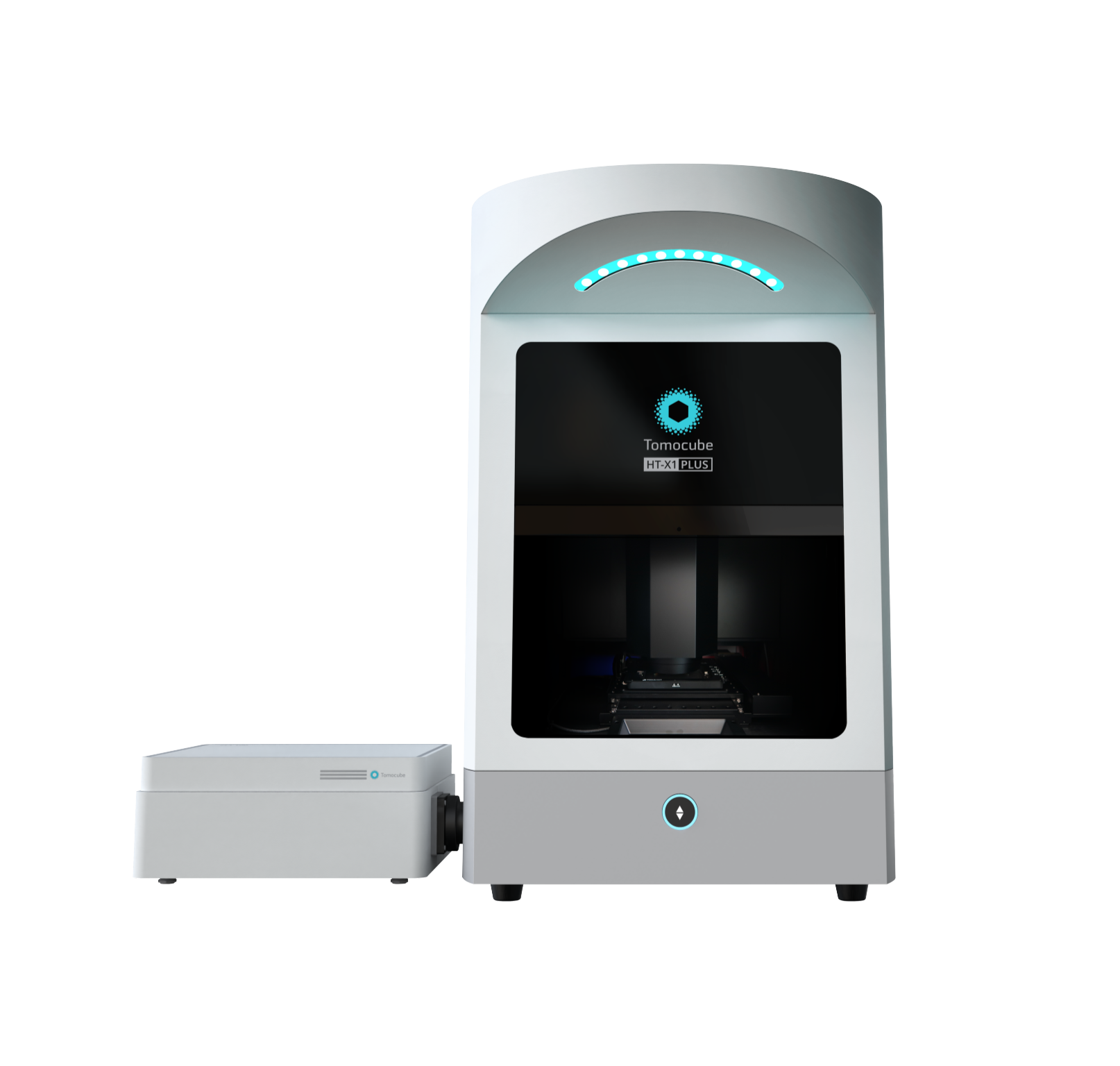
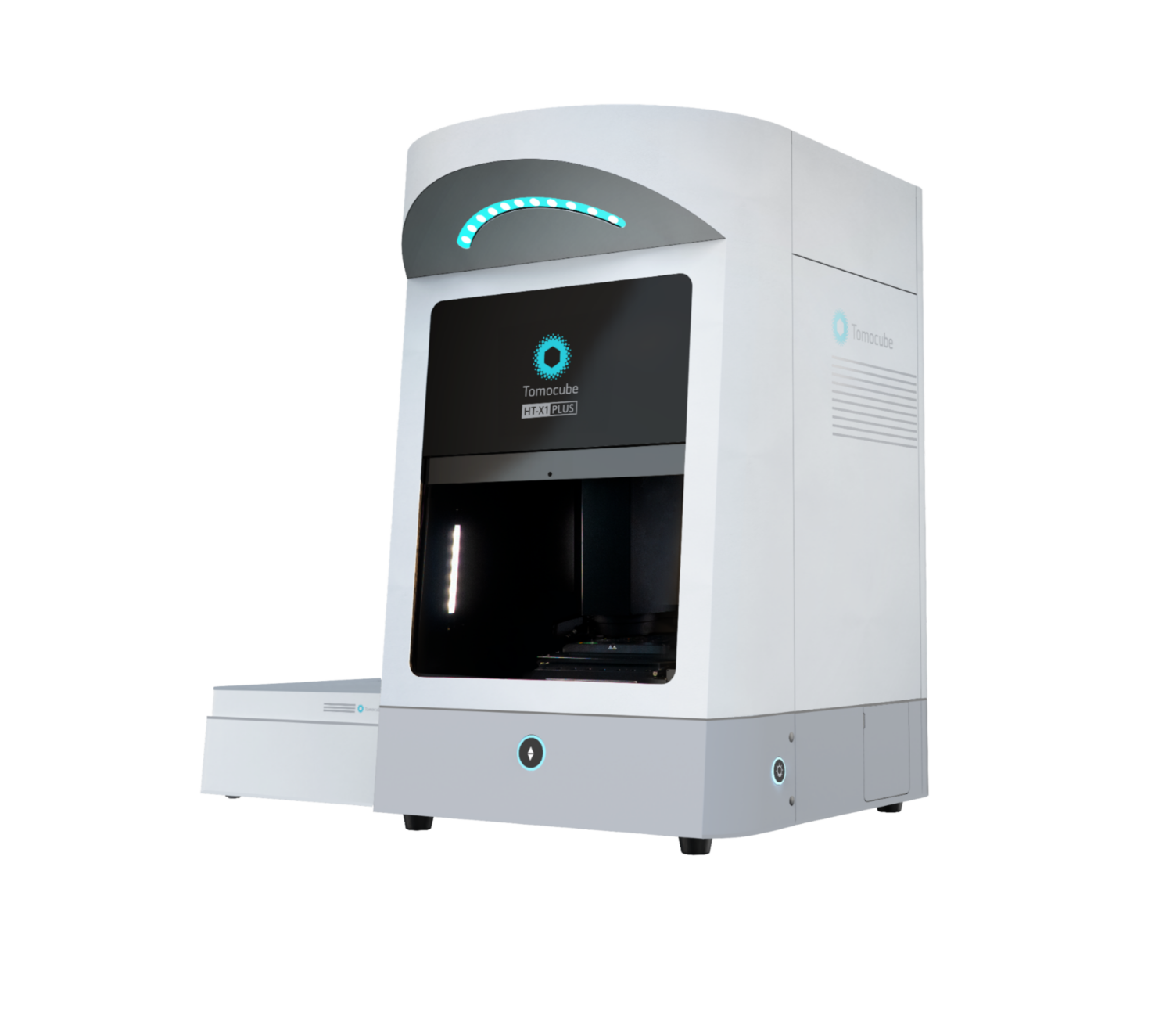
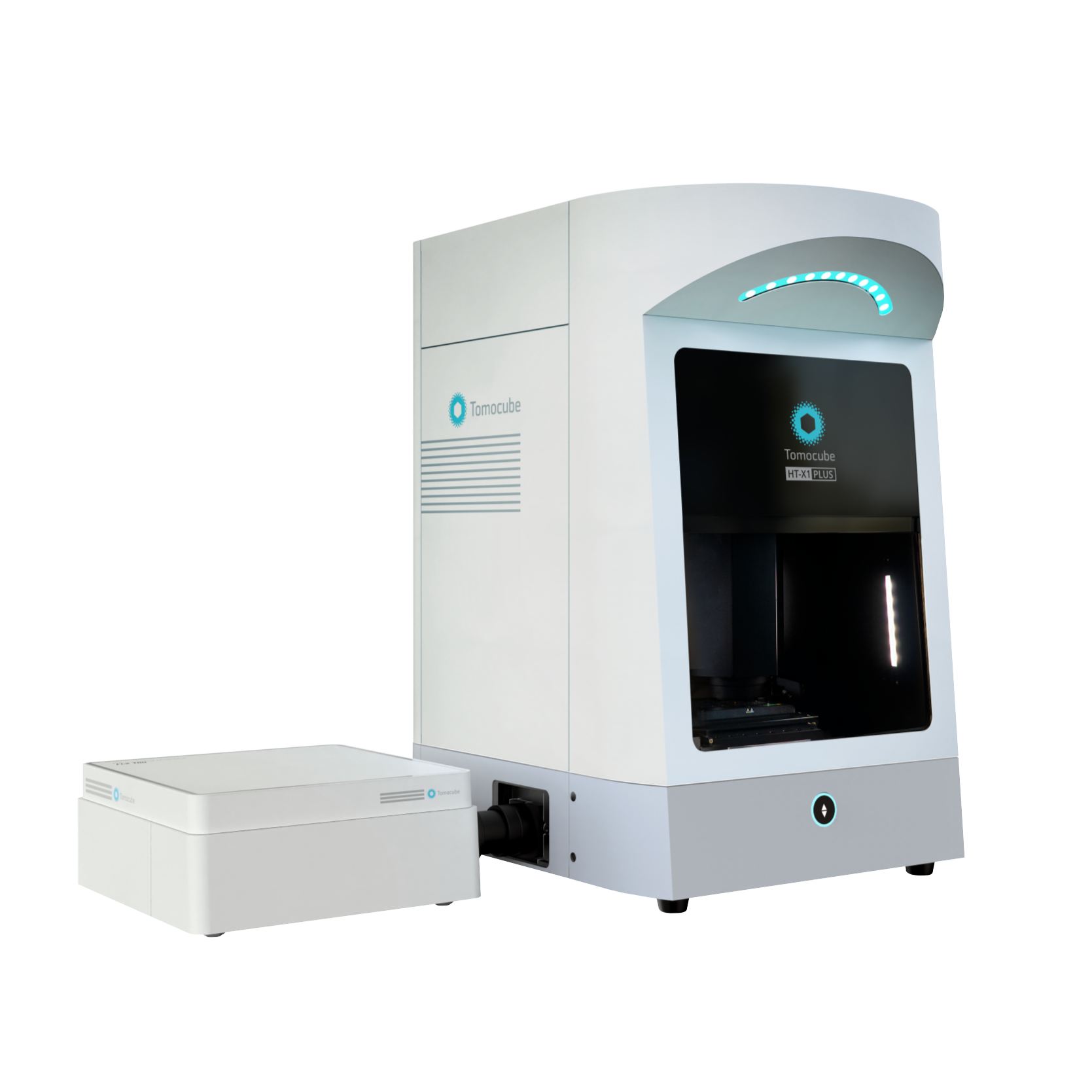
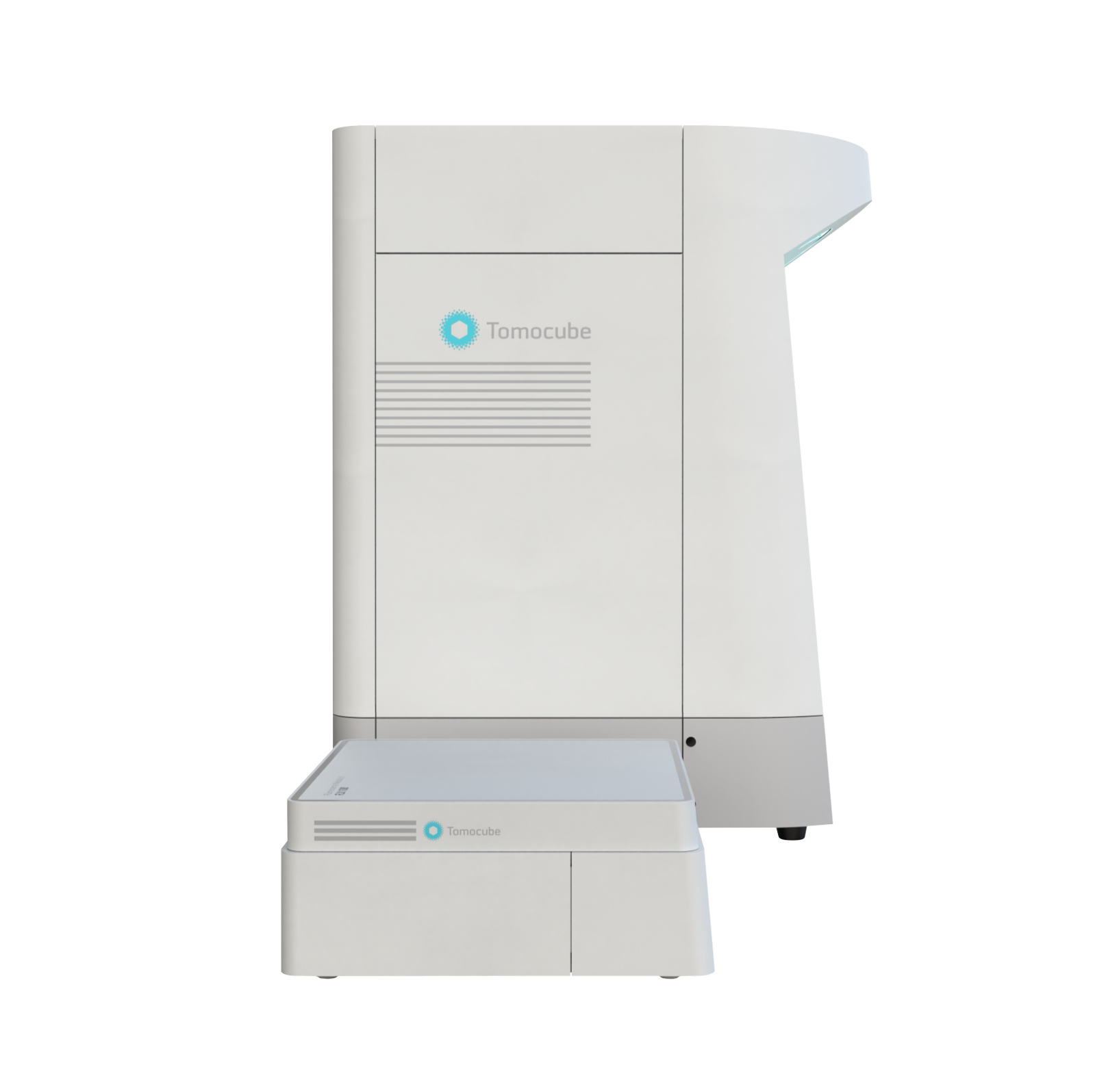
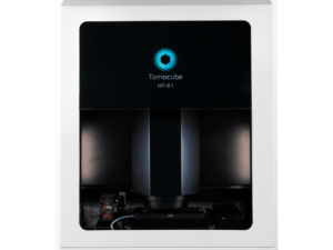
Description
“Equipped with a high-spec camera featuring a 4x larger field of view and significantly reduced acquisition time, the HT-X1™ Plus is perfect for high-throughput phenotypic screening of cells and organoids. Its upgraded correlative imaging capabilities—incorporating an sCMOS-based fluorescence module—enable seamless integration of molecular studies with single-cell-resolution 3D images.
The HT-X1™ Plus extends the reach of Holotomography to an even broader array of challenging specimens, including dense organoids, tissue sections, and fast-moving microorganisms. It is a state-of-the-art Holotomography imaging platform, designed to empower researchers with the precision, efficiency, and reliability needed to drive the future of biological and biomedical discovery.”
Key Features:
- Large Field-of-view
Capture expansive areas without the need for stitching, ideal for large-scale, high-content experiments.
- Faster Image Acquisition
Efficiently acquire high-content images, making it well-suited for high-throughput screening and monitoring dynamic specimens.
- Flexible Choice of Light Source
Customize your imaging with three wavelength options to enhance contrast or improve penetration, tailored to your research needs.
- Combine Advanced Fluorescence
Maximize 3D imaging quality by integrating HT with the sCMOS-equipped fluorescence module, delivering cutting-edge 3D fluorescence imaging
- Wide Preview + Colour Brightfield
Gain deeper insights into tissue section studies with wide preview scan mode paired with correlative color brightfield imaging
User Benefits:
- High Content Screening of Live Cells and Organoids
The HT-X1™ Plus is optimized for high-throughput screening, making it highly suitable for high-content, image-based drug screening research. Featuring a high-performance CXP camera and AI-powered image reconstruction algorithms, the platform excels in both coverage and acquisition speed. Its large field of view measuring 308 μm x 308 μm and rapid 3D scanning capability allows researchers to efficiently analyze an entire 96-well plate in under 30 minutes. This efficiency enables large-scale experiments with unmatched precision and consistency, resulting in faster, more reliable data acquisition that significantly accelerates the drug discovery process.
- High Resolution Imaging of 3D Biological Samples
The new features of the HT-X1™ Plus are especially advantageous for research involving 3D cultures, enabling the detailed investigation of dense organoids and intact tissue sections. The platform incorporates long-wavelength light sources, improving penetration depth and reducing scattering noise, to achieve clearer 3D visualization. Additionally, an advanced 3D reconstruction algorithm further enhances both imaging clarity and precision, delivering superior image quality for 3D biological imaging.
- Enhanced Correlative Fluorescence Imaging
The HT-X1™ Plus offers enhanced multimodal imaging capability with its fluorescence module (FLX™) featuring an sCMOS camera designed specifically for precise signal intensity measurements. The FLX™ module offers high sensitivity to fluorescence, achieving better signal-to-noise ratio (SNR) and shorter exposure times. This allows researchers to obtain biomolecular specificity information from target organelles or fluorescence sensors, even in samples with weak fluorescent signals, such as antibody reactions or hard-to-stain organoids.
- Color Brightfield Imaging for Histological Studies
With the new color brightfield imaging modality and wide preview scan features, researchers can gain deeper insights into tissue section studies. The platform allows for the seamless integration of complex structural data, obtained through 3D optical sectioning, with rich histological information from H&E staining or immunohistochemistry. This integration enhances our understanding of tissue morphology and dynamics, accelerates advancements in clinical pathology and diagnostics, and helps pave the way for the future of personalized medicine.
Technical Specifications:
HT-X1™ Plus Holotomography System Main Unit
Dimensions : 565 (W) × 732 (D) × 921 (H) mm
Weight : 95 kg
Power Supply : 100-240 VAC, 50/60 Hz, 5-3A
Imaging area : 100 mm × 60 mm
Objective lens 40x NA 0.95 air
Objective working distance 180 μm
Condenser lens NA 0.72
Image sensor 20 Megapixels CMOS, CXP-12
Field-of-view : 308 μm × 308 μm
Auto focus : Laser-assisted active sensor
Imaging Modalities : Holotomography, Brightfield (Color), Fluorescence
Supported labware: Dish/Plate/Slide with #1.5 bottom thickness
Wide preview image sensor: Color CMOS
HT-X1™ Plus Holotomography Optics
Light source : LEDs
Illumination wavelength : 444, 520, 660 nm
Axial scan range : 30 – 140 µm
Lateral resolution : 156 – 332nm
Axial resolution : 803 – 1106nm
Minimum acquisition speed : 0.5 sec per images
Fluorescence Module X for HT-X1™ Plus
Dimensions : 434 (W) x 409 (D) x 174 (H) mm
Weight : 25 kg
Light source : LEDs
Excitation filters : 378/52, 474/27, 554/23, 635/18 (nm)
Emission filters : 432/36, 515/30, 595/31, 698/70 (nm)
Filter exchange time : 100 ms
Fluorescence image sensor : sCMOS
Quantum efficiency : 95% (wavelentgh: 580 nm)
Fluorescence light source trigger : 3 channels
Environmental controller
- Model STXG-WSKMXA22B (Tokai HIT)
- Dimensions 151 (W) x 263 (D) x 196 (H) mm
- Weight 3.8 kg
- Temperature setting range Sample temperature: 37 °C
Top heater: 10 °C – 65 °C
Bath heater: 10 °C – 50 °C
Stage heater: 10 °C – 50 °C
Lens heater: 10 °C – 45 °C
10 min to reach 50 °C
- Temperature accuracy Within 0.3 °C
- Humidity control
Heated humidification by the heating bath unit
Recommended water volume: 32 mL
- CO₂ concentration range 5% – 20%
Control method: PID control
Accuracy: ±0.1%
100% CO₂
Input: 0.1 MPa – 0.15 MPa
Output: 160 mL per min
100 – 240 V AC ±10%, 50/60 Hz
- Maximum power consumption
110 W
- Immunology
The immune response is a complex and dynamic process involving intricate interactions among various cells. To gain a comprehensive understanding of this process, it becomes crucial to observe the real time shape and movement of immune cells. In the investigation of immune responses, Holotomography (HT) emerges as a transformative tool, providing unique advantages over traditional imaging technologies.
Holotomography facilitates a detailed exploration of the immune response, allowing for the observation of immune cell shape and movement in three dimensions in real time. Its exceptional ability to instantly capture images of living cells without the need for labeling is an invaluable asset for studying immune responses. This technology also proves instrumental in deciphering processes such as how immune cells recognize and eliminate infected cells.
- 3D biology
Recent advancements in life sciences have prominently featured the development of 3D cell biology. This includes the use of multicellular structures like tissue sections and the creation of 3D culture systems such as spheroids, organoids, and organ-on-a-chip (Microphysiological system). These innovations mark a significant departure from 2D cell cultures, which, while valuable, are inadequate in accurately mimicking the complex spatial cellular organization and tissue dynamics found in vivo.
In 3D biology research, conventional imaging techniques have inherent limitations. The use of exogenous agents for labeling or staining often interferes with cellular functions and compromises data reliability. Additionally, it remains challenges to capture the complete axial structures of thick specimens, such as organoids, or to accurately depict the tissue microenvironment, including dynamic fluid flow, mechanical cues, and tissue-tissue interfaces.
Tomocube Holotomography (HT) is ideally suited for research utilizing 3D cell cultures. The HT-X1 imaging platform offers several key advantages: (1) the ability to capture the complexity of 3D structures, (2) accessibility for non-invasive, live observation of tissue dynamics, and (3) quantitative measurement capabilities suitable for high-throughput applications.
First, HT visualize biological samples by capturing its structural information through light wave, encoding this information onto 2D intensity images, then reconstructing these images into a 3D tomogram for three-dimensional visualization. Through optical sectioning, the HT-X1 enables visualizing depth-specific slices of thick samples and 3D image. Its fast scanning speed and absence of speckle noise are remarkable features in 3D biology applications, ensuring a high-contrast view of the dynamic complexity within 3D models.
Second, HT utilizes refractive index (RI) as image contrast. By minimizing the need for labeling, it preserves cell viability and enables long-term monitoring of cellular dynamics without the risk of photobleaching or phototoxicity. Imaging with HT-X1 allows 3D samples to be observed in their intact forms and without time-consuming, labor-intensive sample preparation protocols. This preserves data reliability and reducing burdens on researchers. Additionally, its fast acquisition speed provides high temporal resolution, capturing rapid cellular events.
Furthermore, HT facilitates quantitative measurement of 3D tissues and organoids, including size, volume, mass, and growth rate. This capability supports comprehensive, longitudinal tracking of the samples’ characteristics and can be leveraged in high-throughput efforts for assessment of drug response.
In summary, Holotomography addresses the limitations of existing imaging techniques by providing high-resolution, label-free imaging of 3D cell cultures. Its noninvasiveness, combined with fast scanning speeds, high-contrast visualization, and quantitative measurements, makes HT an invaluable live cell imaging tool for 3D cell biology.
- Regenerative medicine
Regenerative medicine, a dynamic field at the intersection of biology, engineering, and medicine, aims to restore or replace damaged tissues and organs by harnessing the body’s natural healing processes. In this context, imaging and analysis play pivotal roles in understanding tissue regeneration, optimizing therapeutic interventions, and evaluating treatment outcomes.
The need for advanced imaging and analysis tools in regenerative medicine arises from the complex nature of tissue repair and regeneration processes. Traditional imaging techniques, often limited to two-dimensional views or requiring invasive labeling procedures, may not fully capture the intricacies of tissue dynamics. Meanwhile, Holotomography (HT), as an advanced label-free 3D live cell imaging technology, holds immense promise in regenerative medicine.
Unlike conventional imaging methods, HT provides non-invasive, high-resolution 3D imaging capabilities without the need for exogenous labels or stains. This enables researchers to visualize cellular spatial arrangement, extracellular matrix components, and cellular structures and dynamics in their natural state, preserving cell viability and physiological function.
HT’s ability to quantify cellular features, such as volume, concentration, and dry mass, further enhances its utility in regenerative medicine. This quantitative analysis facilitates the characterization of engineered tissues, evaluation of therapeutic efficacy, and monitoring of tissue development over time.
Moreover, sample reusability in HT imaging is particularly advantageous in regenerative medicine research. By enabling repeated imaging of the same sample without altering its integrity, HT allows researchers to track longitudinal changes in tissue morphology and function. This longitudinal analysis is essential for understanding the temporal dynamics of tissue regeneration, optimizing treatment protocols, and assessing long-term outcomes.
In summary, the integration of HT into regenerative medicine research and clinical practice offers a powerful tool for advancing our understanding of tissue regeneration and enhancing therapeutic strategies. Its high-resolution 3D imaging, label-free, quantitative analysis capabilities, and ability to reuse samples make HT indispensable for visualizing and analyzing complex biological processes in regenerative medicine.
- Cell biology
Unlock the future of cellular research with Holotomography (HT), a cutting-edge imaging technology that transforms our understanding of cell biology. As the foundation of biological science, cell biology holds the key to unraveling the mysteries of life at the cellular level—essential for diagnosing diseases and pioneering new therapies.
Holotomography revolutionizes bio-imaging by offering a high-resolution, label-free, 3D, quantitative solution that leverages refractive index (RI) as an intrinsic contrast mechanism. This cutting-edge approach enables researchers to explore the intricate architecture of subcellular structures—like nuclei, mitochondria, lipid droplets— in fine detail, without relying on potentially harmful stains or labels during long-term observation, a requirement that is often unavoidable in other imaging techniques.
HT’s innovative method safeguards the cells’ natural physiology, eliminating the risks of phototoxicity and photobleaching. By maintaining the intact morphology of live specimens, HT can detect various microstructures without relying on fluorescence markers, which are often lost or degraded during staining and sample preparation processes. This ensures a more accurate and reliable analysis, preserving the true state of the cells under observation.
The benefits don’t stop there. HT offers full 3D visualization, giving researchers the ability to explore cellular structures from every angle, alongside quantitative measurements of cellular properties like dry mass and volume. This wealth of data provides deeper insights into cellular behavior and function, paving the way for breakthroughs in biomedical research.
Holotomography is more than just a tool—it’s a revolution in cell biology, addressing the critical challenges of imaging with unparalleled accuracy and clarity. With HT, researchers are empowered to push the boundaries of discovery, unlocking new potential in the study of life at its most fundamental level.


